Locust Dissection
The Aim
The aim of this dissection was to observe the gas exchange system of the locust. The specific features we were looking for were:
The Aim
The aim of this dissection was to observe the gas exchange system of the locust. The specific features we were looking for were:
- The spiracles, closable holes in the abdomen and thorax of the insect in which oxygen enters the insect
- Trachea, ribbed tubules of chitin which are attached to the spiracles
- Tracheoles, permeable thin tubules attached trachea held extremely close to muscle fibres, which need large volumes of oxygens
- We used sharp objects; scalpels, pins and a pointing needle. These are imperative when doing a dissection so we just have to be careful when using them. If we use scissors make sure to cut away from yourself and always point sharp blades downwards when holding them. We also, when not using them, put the scalpels back in a box to prevent them being left around carelessly
- We also used thin glass cover slips which if we put pressure on would crack which would be quite dangerous. To stay safe we held them in the palm of our hand where they would rest but we wouldn’t hold them.
- Adult locust
- Cork surface
- To attach the pins to
- Dissecting pan
- Scalpel
- Forceps
- Teasing needle
- Monocular
- To provide detailed observation of the locust as spiracles are quite small
- Scissors
- Microscope
- Slide and cover slip
- A watch glass
- To prepare the sample for the cover slide
- Pins
- To ensure precision when cutting the locust it needs to be held down
- Pin the adult locust to the cork surface on its side. Identify using the monocular where the spiracles are situated.
- Prepare the locust for dissection by cutting the wings off and pin some of the legs to the cork surface with the underside of the locust facing towards you
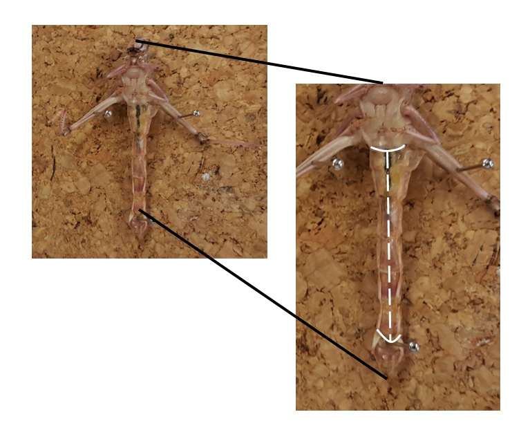
3. Then cut laterally along the centre of the abdomen, then cut longitudinally along the base and the top of the abdomen. These are displayed on the above picture in white. Then pin the scaly flaps you have just formed with your cuts. To help manipulate the flaps into a good observational position you can use the forceps. A picture below explains this step
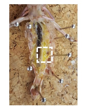
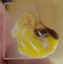
4. From this position you can see many of the larger key features however the tracheoles would be too small to see. Using a scalpel and scissors cut a chunk of the internal organs and put it in a watch glass with a small volume of water. This will clean the organs and make the tracheoles more visible
5. Then place the chunk on a microscope slide and use a cover slip. Then place your prepared slide on the stage of a microscope
5. Then place the chunk on a microscope slide and use a cover slip. Then place your prepared slide on the stage of a microscope
6. Then starting at the lowest level of magnification work up to a good degree of focus and observe the tracheoles that are in-between the muscles and organs
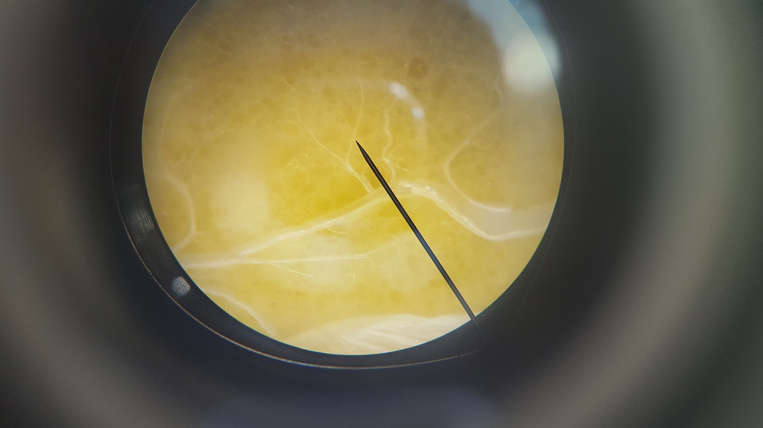
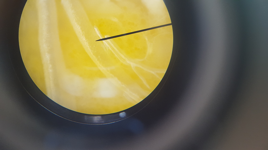
These pictures of the trachea / tracheoles down the microscope, the first picture shows a trachea, in the bottom right corner which branches into smaller tracheoles that are embedded in the yellow clumps of cells. The second picture shows trachea, and if you look closely you can see the ribbed indentations of the chitin. The trachea has chitin so it does not lose oxygen on the way to the tracheoles.
Review
Although this was not a true experiment we still had certain things we were looking for and we did on all the locusts dissected. We were looking and although not measuring directly, we looked at the size comparison of the trachea and tracheoles. I would suggest doing another set of dissections using locusts which have been bred in different oxygen concentrations. I have suggested this after reading an article on a study comparing insect sizes at different oxygen concentrations. They found that insects would grow bigger when the atmosphere had a higher oxygen content, it would be interesting to compare the gas exchange system of all these different locusts at different atmospheric conditions. You could quantitatively compare the size of the different parts of the system.

I think the lack of analysing and recording the spiracles sizes meant our dissection was very observational and did not have much to interpret so using a lens with a ruler in could allow us to measure this in another experiment.
References
http://www.wired.com/2010/11/huge-dragonflies-oxygen/ - article about insect sizes at different concentrations
Comparative anatomy of tracheal system
By: WHITTEN, JM
Annual Review Of Entomology
Pages: 373
DOI 10.1146/annurev.en.17.010172.002105
Published: 1972
Hartung, D. K., Kirkton, S. D. and Harrison, J. F. (2004), Ontogeny of tracheal system structure: A light and electron-microscopy study of the metathoracic femur of the American locust, Schistocerca americana. J. Morphol., 262: 800–812. doi: 10.1002/jmor.10281
Vinal, Stuart C.. “The Respiratory System of the Carolina Locust (dissosteira Carolina Linne)”. Journal of the New York Entomological Society 27.1 (1919): 19–32
Chitin in insects: structure, function and regulation of chitin synthases and chitinases
DOI: http://dx.doi.org/10.1242/jeb.00709
PUBMED: 14610026
