If a picture does not load, refresh the page (f5)
In this, dare I say, practical we were tasked with the duty of created models of meiosis with cookies. This would have to include prophase one, metaphase one, anaphase one and telophase one. But then also document the splitting of the diploids into haploids.
In this, dare I say, practical we were tasked with the duty of created models of meiosis with cookies. This would have to include prophase one, metaphase one, anaphase one and telophase one. But then also document the splitting of the diploids into haploids.
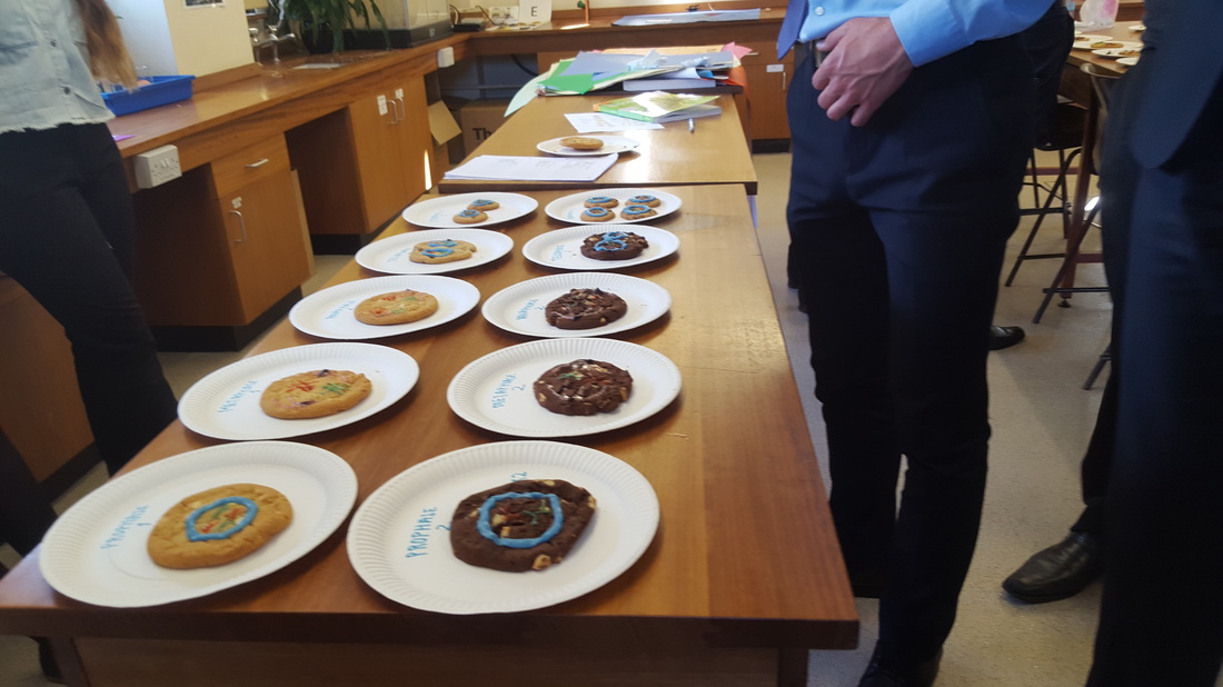
This first picture shows the overall product of another groups more sucessful attempt. Closest to the camera is the prophase of each division. Chocolate chip cookies indicate the meiosis one occuring and double chocolate chip cookies being prophase two. In prophase one the chromosomes condense, this can slightly be seen within the blue ring of icing. The chromosomes are represented by green and red icing. Prophase two is really just the telophase one but is just indicative of how the second stage of meiosis begins.
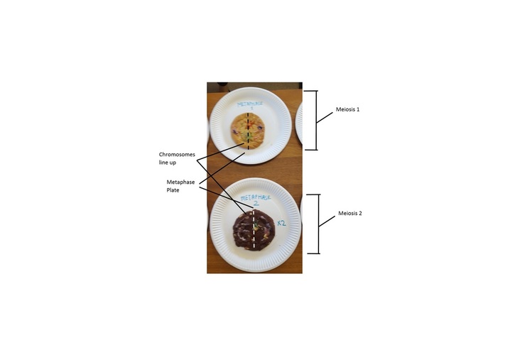
The next picture shows both metaphase one and two. In this the chromosomes have moved to the center, this is highlighted by the dotted lines. The chromosomes are now on the metaphase plate and the centrioles, slightly observable at 3 and 9 o'clock within the cell have moved to opposite poles and spindle fibres have bound to the centromeres of the chromosomes. In metaphase two we can see one of the products of the first stage of meiosis, at this point the chromosomes cross over and the chiasma swap over, this leads to genetic variation. This is shown by the icing has swapped colour at the foot of the chromosome
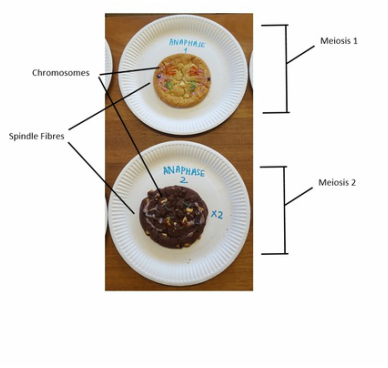
This image shows anaphase one and two. In anaphase one the centromeres are still intact and the pairs of chromosomes have been separated, the chromosomes have been pulled by the spindle fibres they were attached to away to the peripherals of the cell. In anaphase two the centromeres split breaking the chromosome into two chromatids with are half the genetic information. These are moved to the edges of the cell also and like anaphase one the cell begins to split
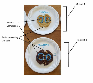
This clip shows telophase one and two. In telophase one and two the cells have begun to split due to a layer of actin around the middle of the cell beginning to contract and cut the cell in have. In stage ones case this means split into to diploids as both nuclear membranes, which have reformed, have got the exact amount of DNA of the parent cell. In telophase two the nuclear membranes only contain chromatids, 23 of the 46 pairs of chromosomes. These chromatids are unique from each other and the parent cell because of the recombination.
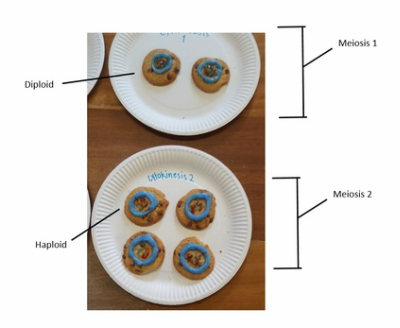
The final picture shows cytokinesis one and two. The final cells are both presented as smaller cookies, although cytokinesis one technically presents two diploid cells so there should be no size difference. However in cytokinesis two the small cookies indicate haploids which contain half the genetic code. An example of this is spermtozoa, which is the haploid male gamete.
Overall I feel presenting meiosis, a concept I originally found counfusing as cookies and creating the models ourself helped benefit my understanding of the concept and process itself
Overall I feel presenting meiosis, a concept I originally found counfusing as cookies and creating the models ourself helped benefit my understanding of the concept and process itself
