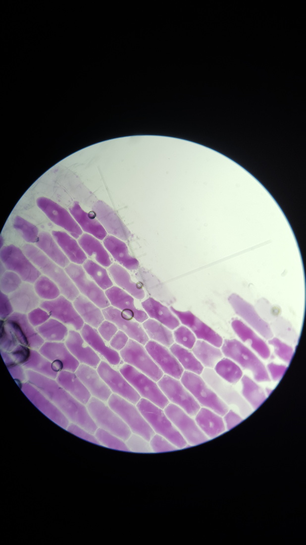
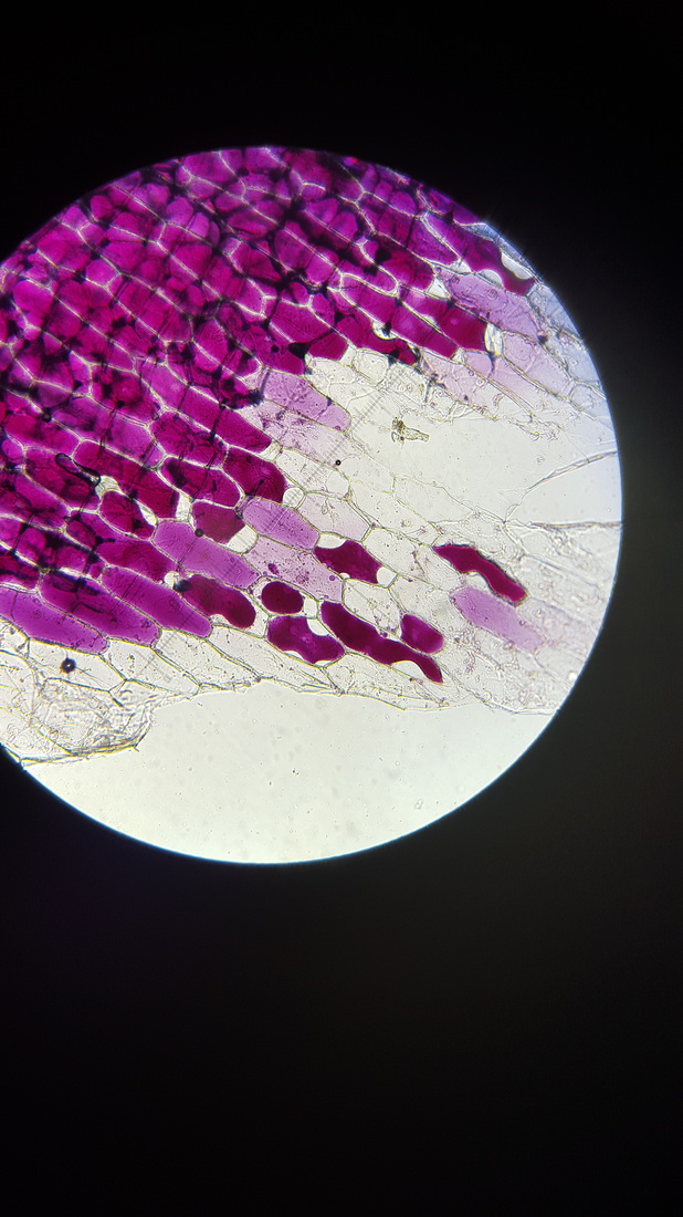
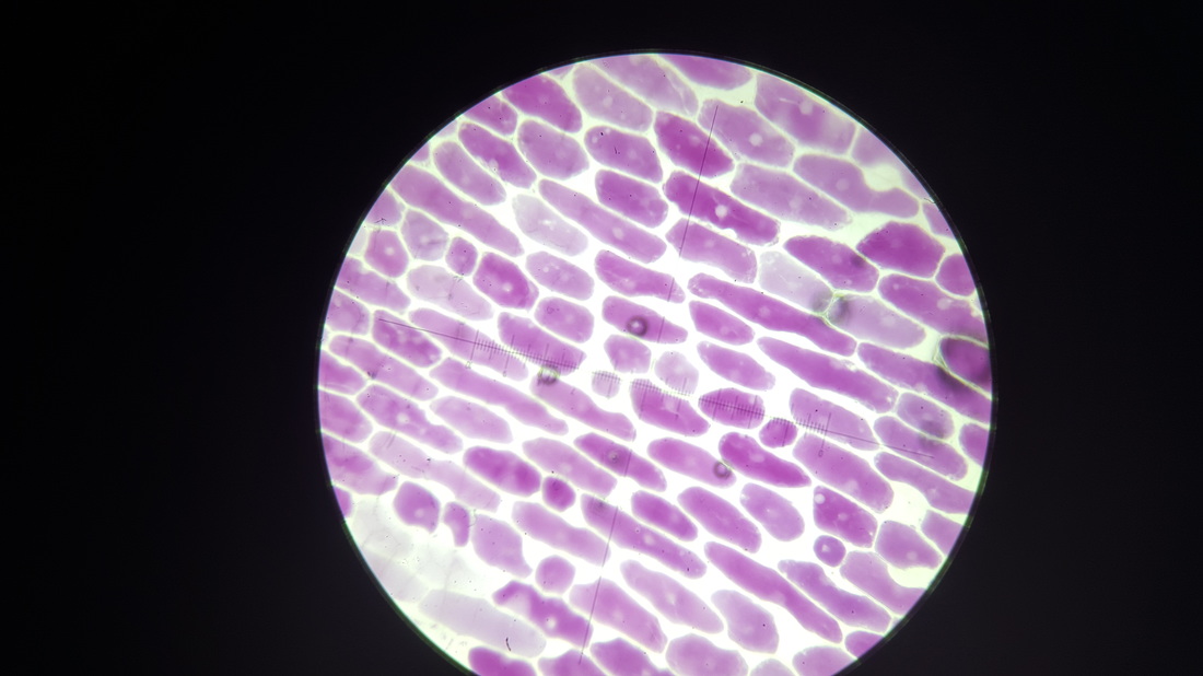
We did this experiment to observe osmosis occurring in cells. Osmosis is the passive movement of water through a partially permeable membrane along the concentration gradient. In a sucrose solution the liquid surround the cells has a low concentration of water. In this case water in the cells will pass into the space around the cells making the red onion cells flaccid. We can see the flaccid cells if the cell membrane is pulling away from the cell wall. A plant cells structure should be rectangular with an even gap between all cells but turgid cells affect structure and make the cells look uneven within the overall structure.
Method
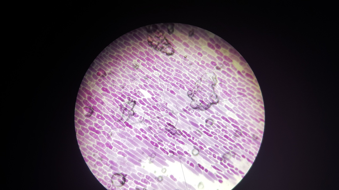
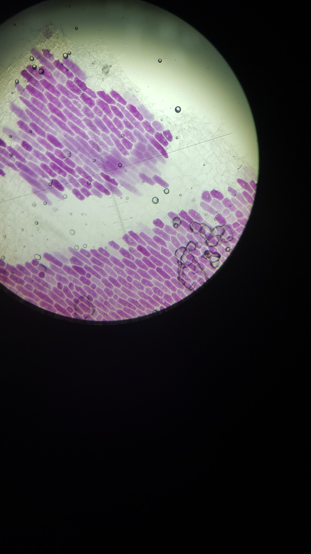
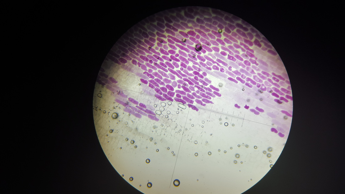
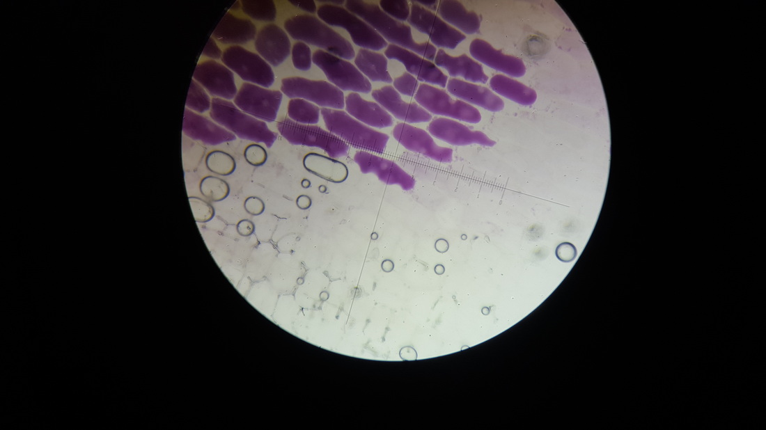
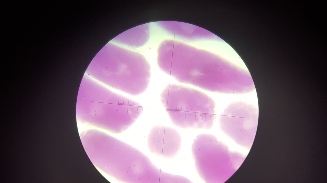
As expected from the chunks of onion skin we saw over time that the cells deformed and changed into an irregular shape.
Method
- Cut a red onion into small segments
- Using a teasing needle cut away the skin away the white part of the onion
- Clear away the residual white parts of the onion leaving just the very thin red skin
- Place the skin on a slide and put a few millilitres of concentrated sucrose solution on the skin
- Gently lower the cover slip onto the slide and soak up excess sucrose solution with filter paper
- Observe the cells over time to see them become flaccid
As expected from the chunks of onion skin we saw over time that the cells deformed and changed into an irregular shape.

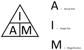
We can calculate the actual size of the cells using the equation above.
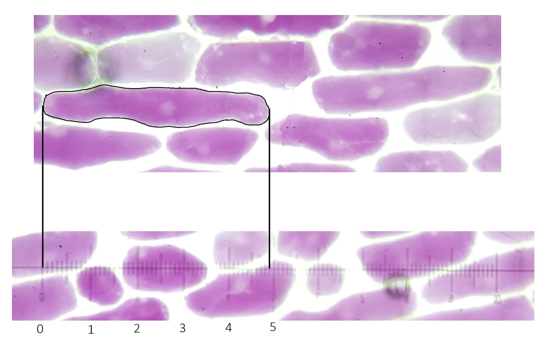
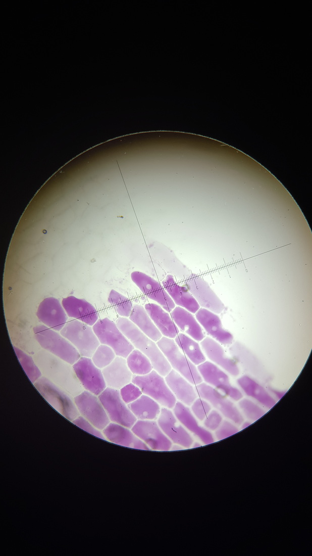
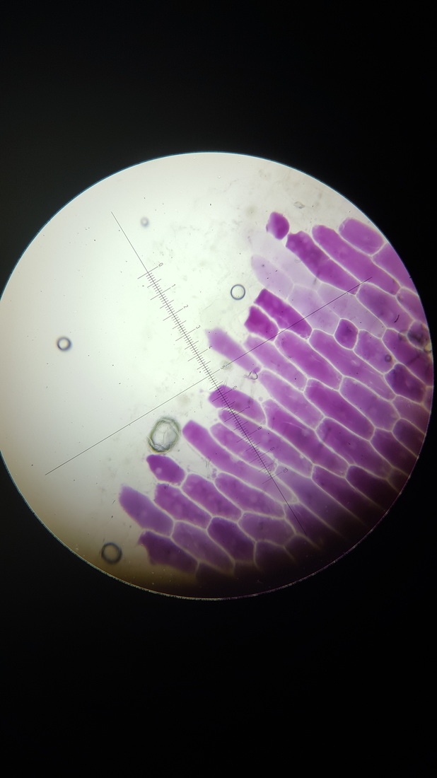
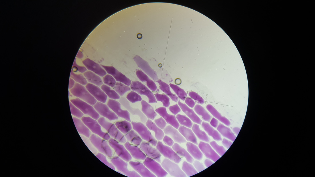
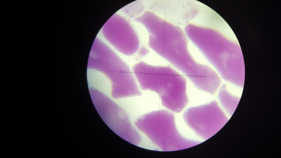
In this case we can work out the magnification by combining the magnification of the eye piece (10x) and the microscope. The microscope will either be 4x, 10x or 40x. The picture we used below is at 40x magnification so in total we have a 400x magnification.
We can also calculate the image size from the graticule in the lens. To ensure that I have correctly compared the size of the cell to the graticule I have rotated it to be parallel to the graticule to accurately see that the cell is 4.8 um long.
Actual size = 4.8 / 400
= 0.0012 um